

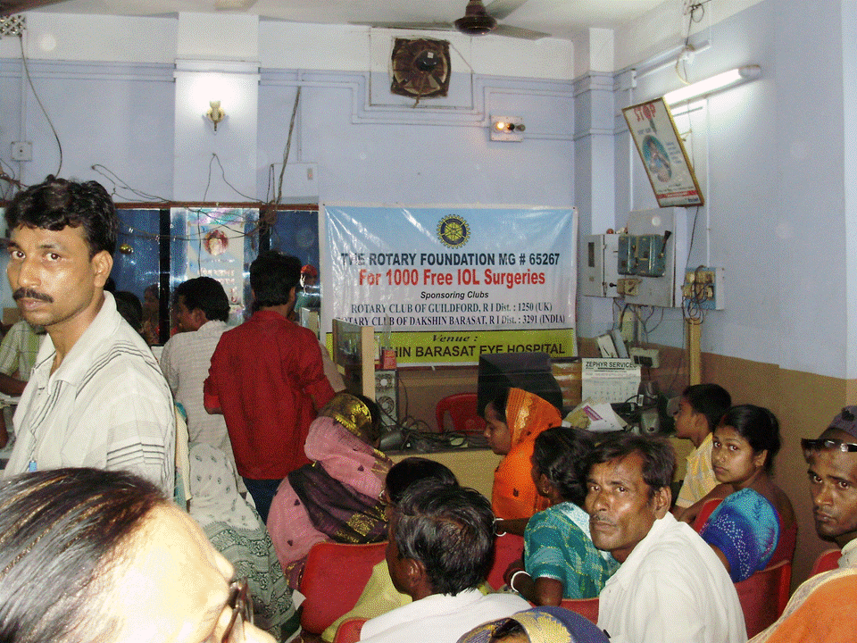
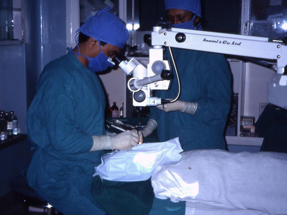


CATARACT OPERATIONS
For new hospitals, existing hospitals who are working towards financial independence, and for hospitals who have already gained financial independence and have subsequently had secondary programmes for further expansion approved, we partner their Trusts in the provision of funding for medical services such as cataract operations.
These tend to be funded in batches of 3000 operations which typically costs less than £40,000.
Grants are often available in support of such provision.
We aim to increase the capacity within Global Sight Solutions' current facilities such that 100,000 cataract operations can be undertaken per year. This is on schedule to be achieved by the end of 2015.
Update...Target achieved on schedule in 2015!
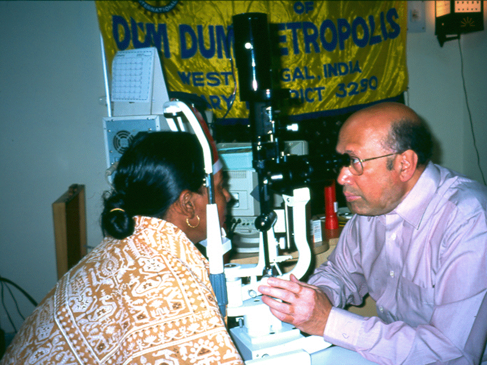
What is Cataract?
In normal circumstances the lens of the eye is clear allowing light rays to pass through it easily and focus on the retina to give clear vision. But when the lens of the eye becomes cloudy it interferes with the passage of light. Hence the image formed in the retina becomes blurred.
Usually cataract occurs in patients above the age of 40. In some instances it can also occur in children. Other diseases such as glaucoma, eye tumours, iritis, and diabetes and also eye injuries may lead up to the formation of cataract. It can also be caused by continuous use of steroids.
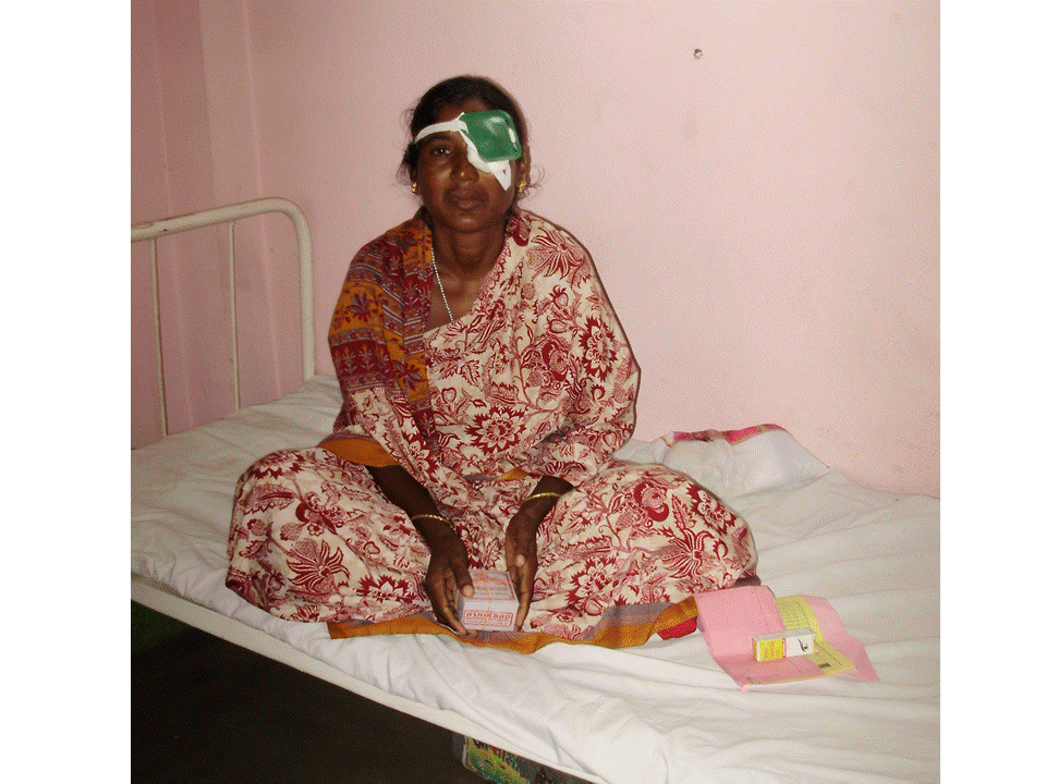
Medicines or glasses wouldn’t be able to cure cataract, the only treatment is surgery where the clouded lens is removed and replaced by an intra-ocular lens. Glasses can be used post surgery to get clear vision.
What is?
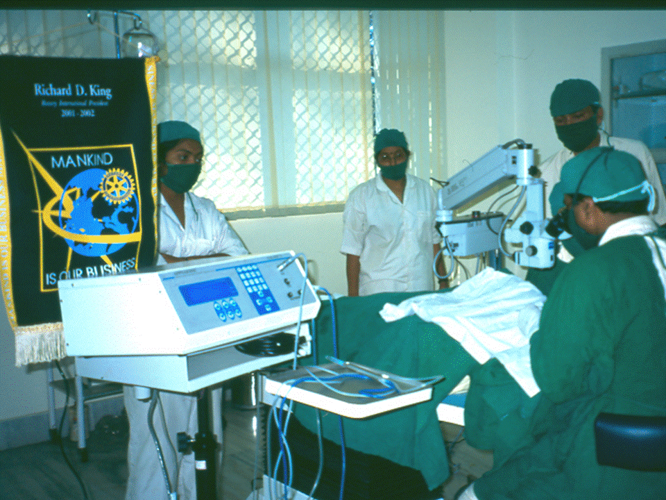
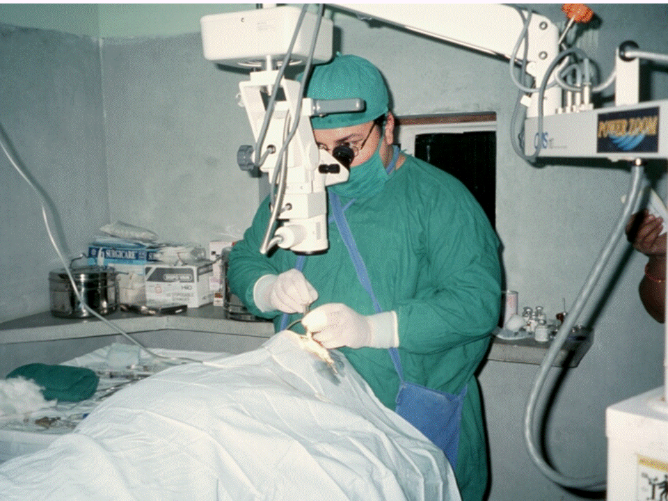
Phaco Surgery and Oculoplasty
Phacoemulsification (Phaco) is a modern cataract surgery in which the eye's internal lens is emulsified with an ultrasonic hand piece and aspirated from the eye. Aspirated fluids is replaced with irrigation of balanced salt solution to maintain the anterior chamber.
Oculoplasty:-
Squint , in layman language means the deviating eye, is a visual defect in which eyes are not in alignment and point in different directions. One eye may look straight ahead, while the other eye may turn inward, outward, upward or downward. Ptosis is a drooping or falling of the upper eyelid. The drooping may be worse after being awake longer when the individual's muscles are tired but that term normally refers to the condition amblyopia. If severe enough and left untreated, the drooping eyelid can cause other conditions, such as amblyopia or astigmatism. This is why it is especially important for this disorder to be treated in children at a young age, before it can interfere with vision development.
A Yag Laser
What is Yag-PI?
-Yag PI is a laser management for patients who may develop glaucoma.
Why is Yag-PI done?
-when a person has not developed glaucoma but falls in the category of glaucoma suspect because of Anterior chamber Angle closure patient goes for YAG-PI.
What is yag cap?
-Yag Cap is the cleaning of the posterior capsule of the pseudophacic patient.
Why is Yag cap done?
-This is done in patients where after cataract surgery, the posterior capsule of the lens opacities in nearly 20% of operated patients.
Glaucoma and Low Vision Clinic
Glaucoma’s are a group of diseases having in common gradual loss of retinal nerve fibres leading to characteristic excavated appearance (called glaucomatous cupping) of the starting point of the optic nerve . This is associated with specific pattern of loss of visual field.
The Low Vision Clinic is a clinic for people who have visual impairment when medical treatment is no longer suitable or is unable to retrieve lost vision. The most common conditions are Age related macular degeneration, Diabetic retinopathy, Glaucoma, untreated cataracts and Nystagmus (continuous uncontrolled to and fro movement of the eyes). However we can see a whole variety of visual impairments.
Paediatric Ophthalmology
Pediatric ophthalmologists focus on the development of the visual system and the various diseases that disrupt visual development in children. Paediatric ophthalmologists also have expertise in managing the various ocular diseases that affect children. Paediatric ophthalmologists are qualified to perform complex eye surgery as well as to manage children's eye problems using glasses and medications. Many ophthalmologists and other physicians refer paediatric patients to a paediatric ophthalmologist for examination and management of ocular problems due to children's unique needs. In addition to children with obvious vision problems, children with head turns, head tilts, squinting of the eyes, or preferred head postures (torticollis) are typically referred to a paediatric ophthalmologist for evaluation. Paediatric ophthalmologists typically also manage adults with eye movement disorders (such as nystagmus or strabismus) due to their familiarity with strabismus conditions.
HFA
The Humphrey Field Analyzer(HFA) is the recognized standard of care for early diagnosis and management of ocular diseases resulting in visual field loss. It is used to confirm that glaucoma has affected the visual function, to evaluate the severity and to monitor progression of the disease.
Cornea & UVEA
The Cornea is the transparent front part of the eye . The cornea, accounts to approximately two-thirds of the eye’s total optical power. In humans, the refractive power of the cornea is approximately 43 dioptres. Any injury or infection of the cornea can lead to damage leading to loss of corneal transparency and thereby disrupting the normal vision.
The UVEA, also called the uveal layer, uveal coat, uveal tract, vascular tunic or vascular layer is the pigmented middle of the three concentric layers that make up an eye. The name is possibly a reference to its reddish-blue or almost black colour, wrinkled appearance and grape-like size and shape when stripped intact from a cadaveric eye. Its use as a technical term in anatomy and ophthalmology is relatively modern.
DCR & DCT
Dacryocystorhinostomy (DCR) surgery is a procedure that aims to eliminate fluid and mucus retention within the lacrimal sac, and to increase tear drainage for relief of epiphora (water running down the face).
A DCR procedure involves removal of bone adjacent to the nasolacrimal sac and incorporating the lacrimal sac with the lateral nasal mucosa in order to bypass the nasolacrimal duct obstruction. This allows tears to drain directly into the nasal cavity from the canaliculi via a new low-resistance pathway. DCT(Dacryocystectomy) Removal of total sac in cases where DCR can’t be performed.